

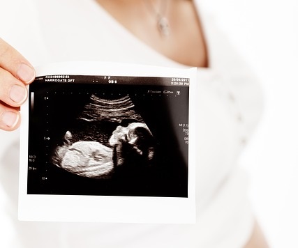
Most of the expecting mothers get an ultrasound check on her to see how her baby looks like. In Sri Lanka most parents-to-be are more eager to know what their baby’s sex is and use this mechanism to check this. No matter what the reason is ultrasounds give you the ability to take a peek at the baby which is growing in your womb (uterus) before it is born.
However, some parents are reluctant to go through this procedure. Some mothers who spoke to BabySpace said that they are afraid that ultrasounds will create health problems in their babies as they are still very mild and vulnerable. Yet, some mothers said that they have done over a couple of ultrasounds on their babies.
‘I have done three on each of my children when they were in the womb and now they are grown ups and they are in perfect health conditions with only a once in a while cold or fever which every other children also have,’ said a mother who shared her thoughts with the BabySpace.
What is ultrasound and what does it actually do? When can a mother do it and what are the conditions a mother should be in to get an ultrasound scan? These questions will be nagging in your mind and in this article we will provide you answers to your questions and help you understand more about the scan.
What really is an ultrasound?
A special machines creates an image of the inside of your body using ultrasound to help you take a look inside it without cutting it open or sending a camera inside your body. These images are more like a small video that appears on the computer screen. In other words ultrasound was introduced in place of x-rays which are known to be harmful to the growing fetus. Also, ultrasound is a ultra high frequency sound wave that creates images accordingly.
Thus it helps you take a look at your baby while it is growing in your womb (uterus). This will provide you the opportunity to see how your baby at that stage looks like and give you important information about the baby such as the size, if the baby is in good health condition such as the working of the heart and if the organs such as brain, kidneys and spine are growing well and even the date the baby is due. It also shows you what sex your baby is, which is a question most parents-to-be and their loved ones wants to know when they are expecting a baby.
Why should I do an ultrasound?
Most people are not very sure of what the ultrasound can do and its benefits. They merely discard the option thinking that most do the scan to see what gender the baby is. But there is a series of benefits to ultrasound that most ignore.
The reasons that you should consider ultrasound are to see if you will have an early pregnancy, or to see both inside and outside the womb. This is done by doing a vaginal ultrasound and also to detect multiple births. To see the placement of the placenta, and to verify the fetal position and breech presentation when it is inside the womb. Also, there are numerous stimulus’s affecting the baby such as diabetes in mothers, ultrasound will monitor the affects and show if there is a threat to the baby, as far as fetal growth and wellbeing is concerned.
Ultrasound is a mechanism that will provide you with these important factors regarding the baby. This way you can get ready or do verification if there is something wrong with the way the pregnancy takes place. But, it doesn’t mean that you should get an ultrasound every time you visit a doctor. Most doctors believe that there is no necessity to get more than two ultrasounds during pregnancy if there is no medical indication.
How is an ultrasound done?
When you visit your doctor for an ultrasound he/she will rub a chilly gel into your belly and then rub a transducer on it. The transducer will detect every slight sound within the womb. Through these sounds the vague images of your baby will be produced in a video screen.
For trans-vaginal ultrasound, a vaginal probe is covered by a rubber condom and lubricant is put on the probe before inserted into the vagina.
There are different types of ultrasound examinations done such as 2D, 3D, and 4D Ultrasounds. There are not all are found in Sri Lanka while not all hospitals in our country provide ultrasounds. Furthermore not all doctors are qualified to conduct Ultrasound scans, and might refer you to a doctor who has the expertise in conducting this.
Types of Ultrasound Scans
Transvaginal scan: used in early stages of pregnancy will be done to see through the vagina and generate sonogram images
Standard abdominal ultrasound: a transducer is used over the abdomen to generate images of the developing fetus
Advanced ultrasound: will focus on a suspected problem in the fetus and will give results using other equipment. This is done by a specially trained fetal medicine specialist with good expertise.
Doppler ultrasound: this measures the slight changes in the frequency of the ultrasound waves
3-D Ultrasound: shows 3-D images of the fetus
4-D Ultrasound: will help you see the facial expression and movements of the growing baby
Fetal Echocardiography: this will assess the heart anatomy and function of the baby and detect any congenital heart defects. This is done by a specially trained fetal medicine specialist with good expertise.
What are the risks in having an ultrasound done?
Up to date many researchers are in debate if long term exposure of the fetus to ultrasound is indeed harmful and cause long term effects. However doctors advise to stick to less than two ultrasounds and only if medically indicated.
- Studies also have shown that 40% of abnormalities are gone undetected by the machine and that it cannot be 100% relied upon.
- As the machine cannot give an accurate finding some parents are stressed and made anxious by the wrong interpretations about the baby.
- Some believe that in rare occasions ultrasounds can cause abnormalities such as learning difficulties, dyslexia, epilepsy or even premature birth and miscarriages. (However it is important to note that these harm haven’t been yet proved scientifically)
There are different types of ultrasound exams which are done in certain periods during the pregnancy. Ultrasounds are normally combined with various other tests such as triple tests, amniocentesis or chorionic villus sampling to certify a diagnosis of Downs syndrome.
Related Article
Ultrasound during different stages of pregnancy